Structure and Functions of Nephron
Dr.A.P.Ganesan
Komarapalayam
Describe the structure and function of a nephron.
Nephron
It is functional and structural unit of kidney.
Each kidney has one million nephron.
They perform excretory, haemostatic and homeostatic functions
Types of Nephron
1. Superficial Nephron
These are situated in outer 2/3 of cortex, They form 85% of the Nephron. These are smaller in size. They function in normal conditions
2. Juxta Metullary Nephron
These are situated in inner 1/3 of cortex, They form 15% of the Nephron. These are larger in size. They function in stress conditions
Structure of Nephron
Nephron Consists of 1. Renal or malpighian corpuscles and 2. Renal tubule
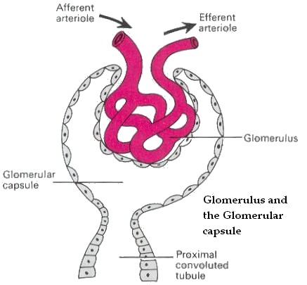
1. Renal or Malpighian Corpuscles
Fount in cortex of kidney .
200 micron in diameter.
consist of two parts A) Bowman's capsule and B) Glomerulus ,
Bowman's capsule
It is the blind upper end of renal tubule . It is invaginated by tuft of capillaries called Glomerulus.
It has two layers namely 1 Parietal layer and 2 Visceral layer.
Parietal layer is made of squamous epithelial cells. It is continuous with epithelial cells of renal tubule .
Visceral layer is made of epithelial cells. This layer is separated from capillary wall by basement membrane which is in contact with endothelium of capillaries.
The Visceral layer, Basement membrane and capillary endothelium together form filtering membrane. Through which filtration occurs. The space between visceral layer and parietal layer is continuous with lumen of renal tubule
Glomerulus
A tuft of capillaries interposed between two arterioles namely afferent arteriole and efferent arteriole.
This capillary tuft invaginates bowman capsule .
The afferent arteriole enters bowman capsule, breaks up into 30 capillary loops called capillary tuft. They do not anastamose. Each loop is enveloped by visceral layer. The capillary loops converge to form efferent arteriole which leaves the bowman capsule close to afferent arteriole
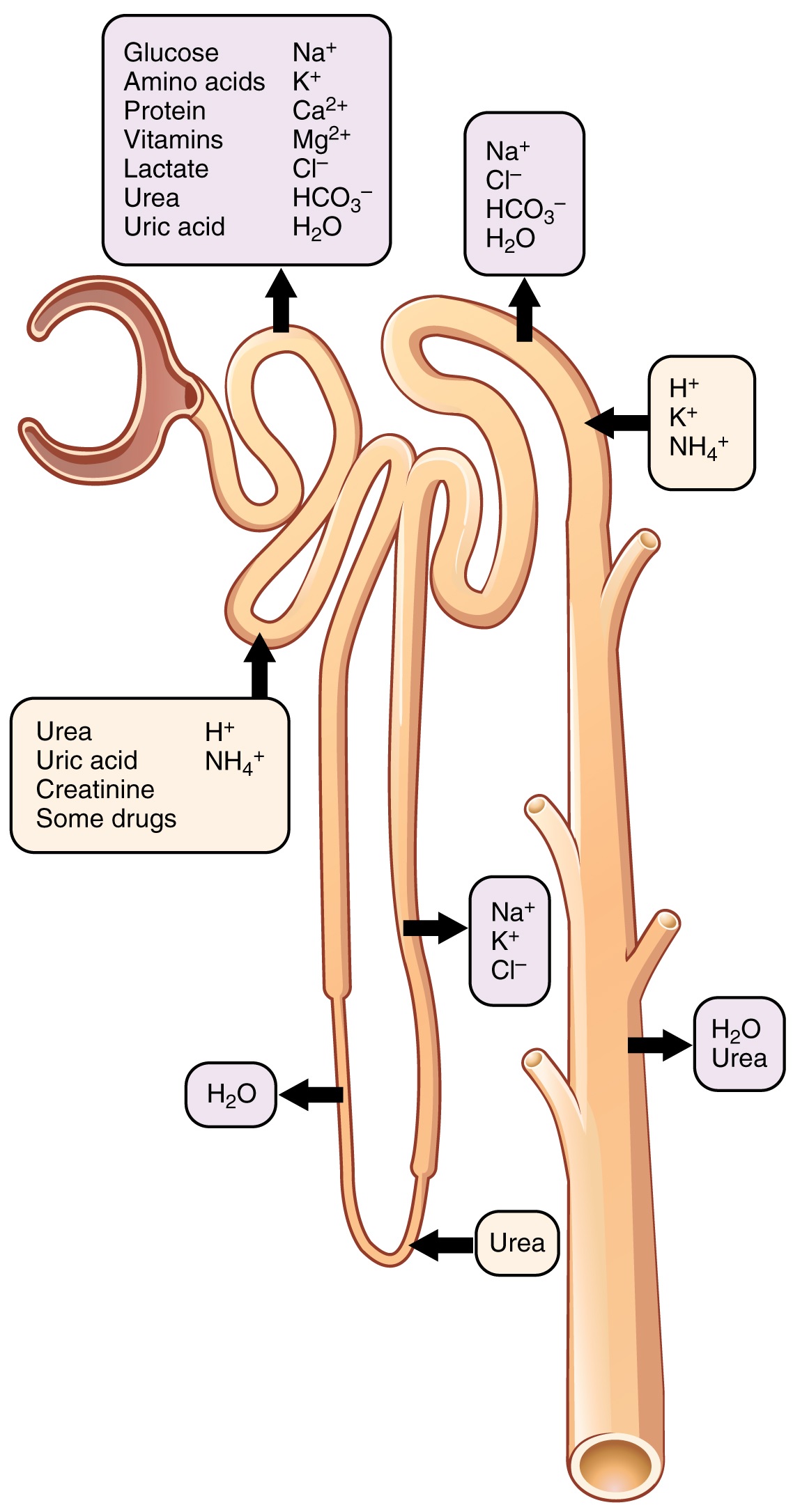
2. Renal tubule
The Renal Tubule consists of the following
- Proximal convoluted tubule
- Loop of Henle
- Distal convoluted tubule
- Collecting tubule
- Duct of Bellini
a. Proxinmal convoluted tubule
A coiled tube, lies close to renal corpuscle, it is 13 to 14 mm in length and 12 to 25 micron in diameter, Its wall is made of single layer of cubiod epithelium, The free surfaces of the calls have microvilli, the cells have mitochondria which supply energy These cells are responsible for reabsorption and secretion. They take reabsorbed water and solutes and deliver the substances, The terminal portion straightens and continues with Distal convoluted tubule
b. Loop of Henle
It is continuation of PCT
U shaped structure
It has descending limb, loop and ascending limb,
The distal 1/3 dips into medulla, It is 12 to 20 mm in length, It is made of flattened epithelium which pulge into lumen.
It continues as distal convoluted tubule.
c. Distal convoluted tubule
It is 4 to 6 mm in length. The initial straight portion passes near bowman's capsule in between afferent arteriole and efferent arteriole, where it becomes convoluted and joins with collecting tubule, It is lined by cuboid epithelium, It is responsible for reabsorption and secretion
d. Collecting tubule
A number of DCTs drain into a single tubule called collecting tubule. This are lined by cuboid epithelium
e. Duct of Bellini
Many collecting tubules join to from Duct of Bellini which opens into apex of papilla or pyramid in the pelvis.
Distal Nephron
DCT, CT, DOB form Distal Nephron It is responsible for final formation of urine
Functions of Nephron
1.Glomerular filtration
2.Tubular Reapsorption
3.Tubular Secretion
4.Formation of New Substances
5.Urine Formation
* * * * * * * * * * * * *